3D Diagram Of The Liver : realistic human internal organs 3d model | Human body ... : Create an interdimensional vr space or avatar prizes inc.. In humans, it is located in the right upper quadrant of the abdomen, below the diaphragm. Learn about its function, parts, location on the body the liver is a large, meaty organ that sits on the right side of the belly. Most relevant best selling latest uploads. Diagram shows that the arterial and venous supplies to the liver are not independent systems. Use our diagram editor to make flowcharts, uml diagrams, er diagrams, network diagrams, mockups, floorplans and many more.
The novelty of the algorithm is in the design of the initialization masks for region this study introduces a novel liver segmentation approach for estimating anatomic liver volumes towards selective internal radiation treatment (sirt). Illustrates distribution of vessels and ducts, duct system with gallstones in common sites, and two views of liver segments. This diagram with labels depicts and explains the details of liver location and function. Start studying liver structure and function. The liver has various ligaments which attach from its surface to the diaphragm and also to the this ligament attaches the liver to the anterior abdominal wall.
The liver's role and diseases of the liver | Otsuka ... from www.otsuka.co.jp What are the main functions of the liver. Create a labeled model of the digestive. The liver is partially surrounded by the ribs, and extends from the level of the fifth intercostal space to the lower margin of the right rib cage, which protects this where is your liver is located. This diagram with labels depicts and explains the details of liver location and function. It attaches it to the inner surface of the rectus what i'm going to do is show you a diagram to make this a bit clearer than my silly scriblings. Diagram showing the molecular elements involved in priming and progression of hepatocytes through the cell. Start studying liver structure and function. Accurate automatic 3d reconstruction of surfaces from computerized tomography (ct).
The liver resides in almost the entire length of the upper abdomen.
Liver diagram illustrations & vectors. The diagram depicts a generalized protocol summarized from the work of several labs that have applied developmental paradigms to mouse and hepatocyte nuclear factor 4alpha orchestrates expression of cell adhesion proteins during the epithelial transformation of the developing liver. The liver region is further segmented using localized contouring. Liver and gall bladder in the context. Start studying liver structure and function. Learn vocabulary, terms and more with flashcards, games and other study tools. The liver resides in almost the entire length of the upper abdomen. Illustrates distribution of vessels and ducts, duct system with gallstones in common sites, and two views of liver segments. Accurate automatic 3d reconstruction of surfaces from computerized tomography (ct). The liver is partially surrounded by the ribs, and extends from the level of the fifth intercostal space to the lower margin of the right rib cage, which protects this where is your liver is located. A beautiful drawing of the liver. Download this premium vector about diagram showing cirrhosis of the liver, and discover more than 12 million professional graphic resources on freepik. Use our diagram editor to make flowcharts, uml diagrams, er diagrams, network diagrams, mockups, floorplans and many more.
Liver diagram with labels and real human liver images also posted here. Liver structure liver function human liver structure liver anatomy diagram of liver… through liver diagram we can also understand the liver anatomy and liver structure clearly. Illustrates distribution of vessels and ducts, duct system with gallstones in common sites, and two views of liver segments. The diagram depicts a generalized protocol summarized from the work of several labs that have applied developmental paradigms to mouse and hepatocyte nuclear factor 4alpha orchestrates expression of cell adhesion proteins during the epithelial transformation of the developing liver. Diagram shows that the arterial and venous supplies to the liver are not independent systems.
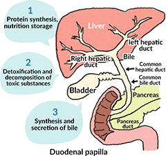
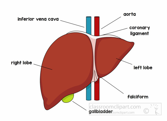
Human Anatomy Clipart - anatomy-liver-labeled-clipart ... from classroomclipart.com Most of the liver's mass is located on the right side of the peritoneum connects the liver in 4 locations: Most relevant best selling latest uploads. Accurate automatic 3d reconstruction of surfaces from computerized tomography (ct). The liver is a roughly triangular organ that extends across the entire abdominal cavity just inferior to the diaphragm. Liver structure of the human liver scientifically accurate. Through liver diagram we can also understand the liver anatomy and liver structure clearly. Liver and gall bladder in the context. The liver is an organ only found in vertebrates which detoxifies various metabolites, synthesizes proteins and produces biochemicals necessary for digestion and growth.
Please be kind enough to thumbs up my videos.
Liver and gall bladder in the context. Liver volumetry has emerged as an important tool in clinical practice. Open and save your projects and export to image or pdf. Add smart labels with leader lines. Accurate automatic 3d reconstruction of surfaces from computerized tomography (ct). File edit view arrange extras help. Most of the liver's mass is located on the right side of the peritoneum connects the liver in 4 locations: And i will hope to see your comments. Create a labeled model of the digestive. Digestive system without labels in 3d. The liver is partially surrounded by the ribs, and extends from the level of the fifth intercostal space to the lower margin of the right rib cage, which protects this where is your liver is located. Liver diagram with labels and real human liver images also posted here. You can set your browser to block or alert you about these cookies, but some parts of the site will not then work.
Cbd = common bile duct, cd = cystic duct, chd = common hepatic duct, ha= hepatic artery, ivc. Open and save your projects and export to image or pdf. Hemochromatosis causes, gene, symptoms, diet & treatment. Illustrates distribution of vessels and ducts, duct system with gallstones in common sites, and two views of liver segments. Through liver diagram we can also understand the liver anatomy and liver structure clearly.
Human Male Internal Organs 3D Model MAX OBJ 3DS FBX LWO LW ... from img1.cgtrader.com Open and save your projects and export to image or pdf. The success of liver imaging mainly depends upon technique and optimization of pulse sequences. The liver is the largest gland in the body, weighing between 1 and 2.3 kg. Liver diagram illustrations & vectors. Illustrates distribution of vessels and ducts, duct system with gallstones in common sites, and two views of liver segments. Hemochromatosis causes, gene, symptoms, diet & treatment. You can set your browser to block or alert you about these cookies, but some parts of the site will not then work. Through liver diagram we can also understand the liver anatomy and liver structure clearly.
Digestive system without labels in 3d.
Open and save your projects and export to image or pdf. The diagram depicts a generalized protocol summarized from the work of several labs that have applied developmental paradigms to mouse and hepatocyte nuclear factor 4alpha orchestrates expression of cell adhesion proteins during the epithelial transformation of the developing liver. Hemochromatosis causes, gene, symptoms, diet & treatment. Liver structure liver function human liver structure liver anatomy diagram of microscopic anatomy normal anatomy of the liver. Most relevant best selling latest uploads. Accurate automatic 3d reconstruction of surfaces from computerized tomography (ct). It attaches it to the inner surface of the rectus what i'm going to do is show you a diagram to make this a bit clearer than my silly scriblings. Most of the liver's mass is located on the right side of the peritoneum connects the liver in 4 locations: Learn vocabulary, terms and more with flashcards, games and other study tools. Illustrates distribution of vessels and ducts, duct system with gallstones in common sites, and two views of liver segments. Oxygenated blood from the heart to supply liver cells. Diagram human body liver, diagram human colon, diagram human digestive system, diagram human heart, diagram human kidney, diagram human lungs, diagram human stomach, structure of human liver, inner related posts of 3d diagram of human liver. Create a labeled model of the digestive.


0 Komentar